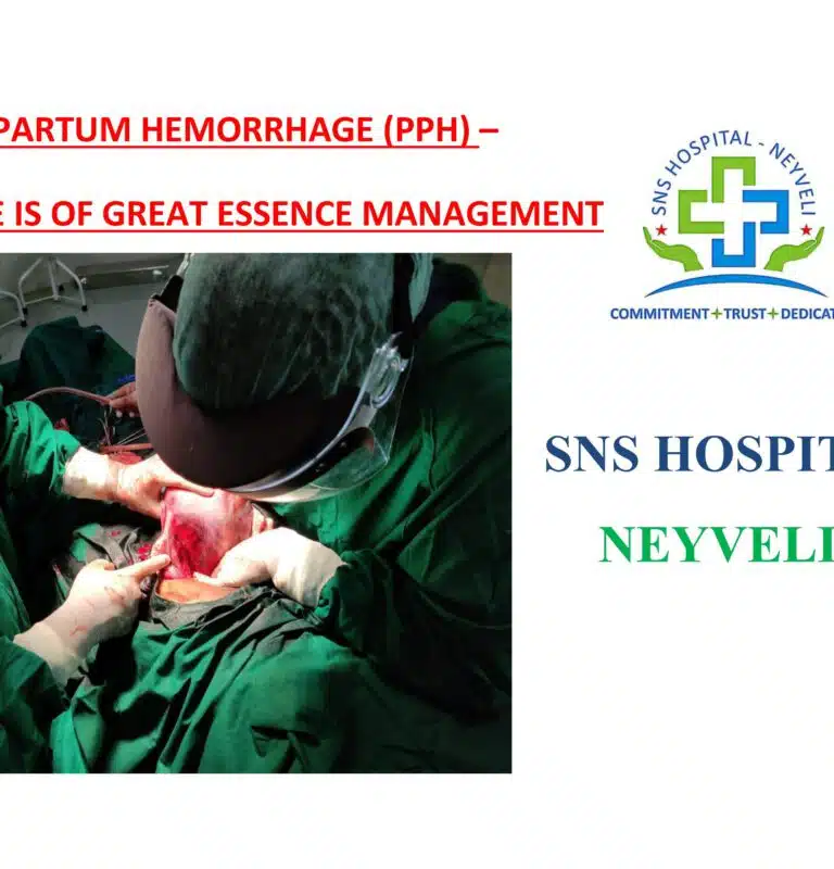
POSTPARTUM HEMORRHAGE (PPH) – “TIME IS OF GREAT ESSENCE MANAGEMENT
Peripartum hemorrhage remains one of the main causes of maternal mortality for decades worldwide.
A key element of the hemostatic management is the development of standard operatingprotocols combining surgical as well as medical and hemostatic treatments depending on the cause and severity of bleeding.
The presentation comprises the significance of using newer uterotonics, tranexamic acid, blood products such as FFP platelets , PRBC .
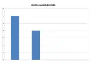
In addition :the significance of antenatal evaluation and preparedness, vigilant monitoring and quick decision making in dealing with obstetric haemorrhage goes a long way in reducing morbidity.
DISCUSSION:
The three take away messages
PPH is any bleeding >500ml on mean within 24hrs of birth .
But it is recommended to be in individualizedbased on the antenatal risk factors andgeographical factors the women belongs to .
The first step of PPH is to recognize that.
PPH is not a diagnosis but it’s a clinical sign of an underlying condition that by itself requires diagnosis.
Though not predicable the major morbidities and mortalities caused by PPH is entirely preventable only by Vigilant Monitoring of not only High Risk But Also Low Risk women and the swiftness and certainty of the team is the greatest predictor of the outcome .
STANDARD OPERATING PROTOCOL - (PPH)
It is highly recommended that every obstetric department develop SOP adopted to its location ,infrastructure and logistics.
SOP should clearly aim to identify PPH at the earliest ,have an escalating mode of management ,draft transfusion protocols checkout the short term and long term mobilities, derive strong consensus for high risk obstetrics.
Assessing biochemical parameters during early labour in all high risk women .
Prophylactic uterotonics .
Cross matching samples.
AMTSL.
Our hospital prefers – inj carbetocin (better shelf life )100ug in during delivery of baby .
Prior information to from blood bank.
Obstetrician and paramedical team on site.
Experienced anesthetist on call.
“TIME IS OF GREAT ESSENCE MANAGEMENT”
A difficult situation of antepartum and postpartum hemorrhage is an enormous source of stress for the team and the patient as well.
Here we highlight the two significant cases..
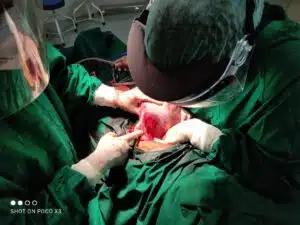
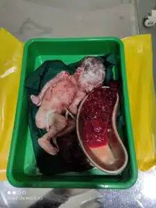
CASES-1
30years | G2P1L1 | Pre caesarean delivery | term /Anemia treated with iron sucrose antenatally / late onset severe preeclampsia ( diagnosed a week back ) / |admitted ( first visit ) on 14/05/2021 @ 8.00 Am with C/O bleeding P/V diagnosed ---ABRUPTION with DI C – Grade IV (with IUD & HELLP ) UNDERWENT laparotomy AT 08.30 AM – intrauterine – dead fetus delivered and retroplacental clots-800gm removed / couvelaire uterus.
Packed RBC and FFPS’AND Platelets transfused .
Postoperatively Patient had central serous retinopathy managed conservatively with CT and opthal opinion
DISCUSSION -
Early intervention in pre eclampsia would have prevented morbidities
Pre eclampsia and anemia is a dreaded combination
It takes just few hours for a pre eclampsia women to land up in HELLP / DIC /IUD – so a vigilant monitoring and stringent protocol is absolutely essential.
CASES-2
G2P1L1 | Pre FTND |booked and immunised outside/ admitted in active phase of labour @ 03.15 pm ( second visit)with thick meconium ,labour progressed well deliverd safe @ 04.35 pm .
Interestingly we found the OS closing within 5 mins of delivery without expulsion of placenta DURING THIRD PHASE OF LABOUR
Patient taken up for vaginal exploration and manual removal of placenta under shout GA @ ---The ends of placenta could not be reached, only the few cotyteledons were palpable on the left cornual end of the uterine cavity.
Decieded for emergency laparotomy @_05.30 pm with high risk consent .
INTRAOP FINDINGS :
BICORNUATE uterus with a communication -placenta in the left horn –at about 500ml of clots in the right horn.
DISCUSSION
A previous normal delivery doesn’t necessarily rule out uterine anomalies
All retained placentae need not be given 30 min separation time.
Uterus is a wonder organ as always - in this case we saw the cavity closes as soon as its empty.
Need not panic about the closing os – understanding the basic physiology would help in diagnosis.
AS ALWAYS KNOWN- MANY UTERINE ANOMALIES ARE DIAGNOSED PERIPARTUM .
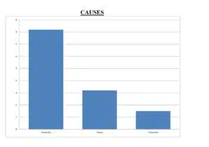
During Dec 2020 –Nov 2021 , we encountered eleven cases of postparatum hemorrhage
each with different etiology - constitutes incidence of 10% of deliveries .
SEVERITYCAUSES
MAJOR PPH - 9 , MINOR PPH - 2ATONICITY-9, TISSUE -1, THROMBIN -1
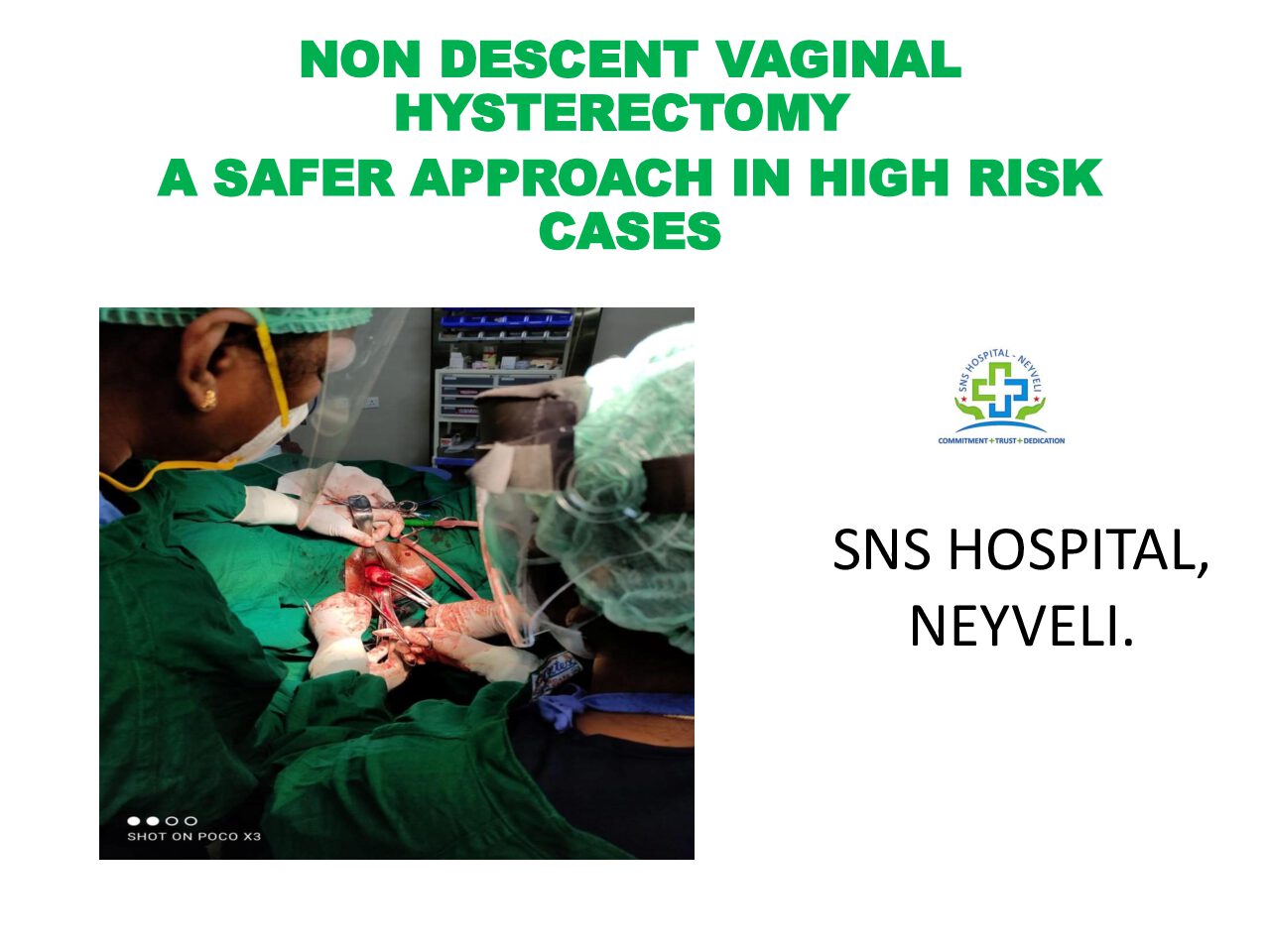
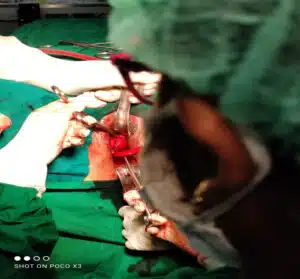
Hysterectomy is one of the common gynecological surgery performed through abdominal/ vaginal /laparoscopic routes. Vaginal techniques have been practiced for centuries, but has been outdated especially in recent years after the practice of Laparoscopic hysterectomies. This is due to lack of training and enthusiasm among evolving gynecologist and due to misconception that abdomen route is safer and easier.
Vaginal route is the least invasive route of all, utilizing a naturally existing anatomical orifice .
The ease and convenience offered by an abdominal incision have led to the preponderance of abdominal hysterectomies.
However proper selection of patient is a critical factor is determining the success of vaginal procedures.
Lack of expertise and curve in learning the technique has major impact on choosing the route of hysterectomies.
In our center hysterectomy is performed by Laparoscopic, Abdominal and vaginal routes.
However we prefer the later route as we find it less time consuming and cost effective for patients.
CASE DISCUSSION - I:
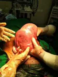
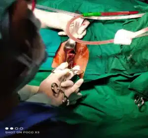
48years Mrs. S , Heavy menstrual bleeding -6 years, Fibroid uterus 14 weeks - 5 cm Intra manual fibroid with Adenomyosis.
Patient was a K/C/O systemic Lupus erythematosus on immunosuppressants.
With H/O lung involvement four years back for which she underwent tracheostomy.
She had several risk factors like hypertension, Renal impairment secondary to SLE/morbid obesity.
We opted for trial vaginal Hysterectomy as even Laparoscopic on abdominal route was risky often this patient.
Patient underwent vaginal Hysterectomy with multi disciplinary perioperative case and was discharged 4 th post operative day.
CASE DISCUSSION - II:
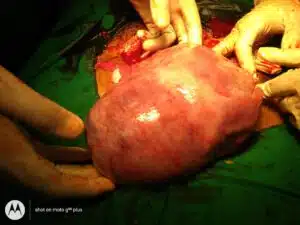
44 years Mrs. X, A P2L2 / Sterilised.
K/C/ - Fibroid Uterus, submucosal with 3cm fibroid polyp protruding through the os.
With C/O - Heavy menstrual bleeding and pain 1yrs.
P/V - Uterus 12week mobile, VTT positive. Underwent vaginal Hysterectomy for non descent Uterus.
Patient discharged on II POD.
DISCUSSION - DOCTORS CORNER:
NDVH is a Passion of Gynec Surgeon.
Vaginal route is the safest and most cost effective route because of shorter hospital stay,
No visible scar .
No risk of General Anesthesia hence can be used in cases with Cardio Pulmonary complications where Laparoscopic route is difficult.
More time Saving is experienced hands. Laparoscopic route is associated with increased operating times and risk injuries.
Quick recovery and early discharge.
Using single clamps is easier in difficulty cases with decreased space.
DIAGNOSIS - GDM ON MEAL PLAN WITH FETAL CARDIAC DEFECTS
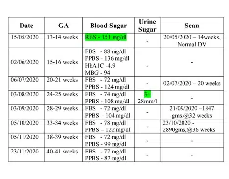
26 yrs – Primi/ Mrs. R 25/F, accountant by occupation , booked elsewhere , LMP-10/02/2020, EDD - 17/11 2020/ First visit to SNS Hospital on 07/10/2020 @ 35 weeks. Known case of GDM on Meal plan since three months of pregnancy / she was under vigilant monitoring of blood glucose , maternal wt gain, Bp, monitoring throughout the pregnancy.
The plasma Glucose levels seems apparently well under control.
Her serial growth scans showed EFW - <50th percentile.
Underwent cesarean delivery on -18/11/2020 for Dystocia of Labour, and delivered Girl – 4.00 kg. Early neonatal and postnatal period uneventful. No episodes of hypoglycemia in the baby. Maternal Blood glucose within normal limits.
CARDIAC screening of the baby was done - is fourth week of life. Showed -
> Acyanotic congenital heart disease.
VSD - 5mm - muscular non -
restrictive.
ASD - 4mm - L-R shunt
PDA - 2.5mm - L-R shunt. Baby underwent corrective surgery and is thriving well.
Points to ponder - Doctors` corner:
Even one marginally higher blood glucose - (151mg/dl - 13weeks) in the earlier trimester -though we name it as GDM - Should, arouse a suspicion of embryonic defects.
All the parameters were apparently under control throughout pregnancy, with meal plan - yet the Fetus had undergone the insult at the very earliest stage - should we redefine the terminology - GDM?
How far does the meal plan take care of the Intra uterine fetus - is Questionable.
Defining any single abnormal blood glucose values as diabetesmelitus complicating pregnancy - like what has been done in this case helps us to keep a close watch.
Asian ethnicity being a high risk for Diabetes complicating pregnancy - we see a tremendous increase in the incidence of number of pregnancies complicated with Diabetes.
Strict monitoring and control of blood glucose helps us prevent late intra uterine complications (sudden fetal death) intrapartum and early neonatal complications, not reliable is preventing embryonic insults though. Which says the pathogenesis of congenital abnormalities seen in babies born to diabetic mothers might be because of cytopathic effect of maternal immune system.
ECTOPIC PREGNANCY OCCURS WHEN A FERTILIZED EGG IMPLANTS OUTSIDE THE UTERINE CAVITY. THE MOST COMMON SITE BEING FALLOPIAN TUBE. ECTOPIC PREGNANCY IS ONE OF THE LEADING CAUSE OF MATERNAL MORBIDITY DURING FIRST TRIMESTER.
CASE STUDY 1:
DIAGNOSIS – TUBAL ECTOPIC PREGNANCY WITH TUBAL LEAKAGE AND PELVIC HEMATOCELE
CASE HISTORY
23 YRS MRS A PRESENTED TO THE OPD ON 26/02/2021 @ 11 .00 AM WITH PAIN – 4 DAYS WITH C/O LOOSE STOOLS –TREATED ELSEWHERE WITH ANTISPASMODICS. HER LMP – 18/2/2021. (PREVIOUS MENSTRATION 7/1/2021).
MANAGEMENT
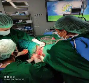
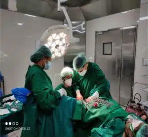
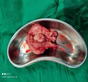
URINE GRAVINDEX WAS DONE WITH SUSPICION FOR TEN DAYS OF AMENORRHEA PRECEDING THIS CYCLE – FOUND POSITIVE AND ULTRASOUND SCAN DONE SHOWED ECHOGENIC MASS 4 * 4 CM LEFT ADNEXA, FREE FLUID + BETA HCG – 5550 Iu/ml. PATIENT WAS PALE WITH HB – 6.8 gm/L. LAPAROTOMY DONE WITH ONE UNIT OF PACKED CELL DONE AT 1.00PM AND SHOWED HEMOPERITONEUM WITH 100 ML OF BLOOD STAINED FLUID ,PELVIC HEMATOCELE – 200gm OF CLOTS, LEFT AMPULLARY PREGNANCY – 4x4 cm. LEFT SALPINGECTOMY DONE AND CLOTS REMOVED, PREITONEAL WASH DONE.SPECIMEN SENT FOR HISTO PATHOLOGY . BLOOD TRANFUSIONS WAS DONE. POSTOPERATIVE PERIOD UNENENTFUL AND PATIENT WAS DISCHARGE ON D3.
DISCUSSION
TUBAL LEAKAGE IS THE MOST USUAL COURSE OF AN ECTOPIC PREGNANCY.
PROCESS TO PERITUBAL HEMATOCELE / PELVIC HEMATOCELE / ON TUBAL ABORTION.
BY TUBAL ABORTION IS MEANT EXTRUSION OF THE OVUM FROM FIMBRIAL END. (COMMON DIAGNOSIS).
10% CASE GO FOR TUBAL RUPUTURE.
PELVIC HEMATOCELE – RECTAL DISCOMFORT.
WHAT THIS CASE TEACHES US -
THE SIGNIFICANE OF HIGH DEGREE OF SUSPICION.
ITS IMPORTANT TO CONSIDER ANY BLEEDING FOLLOWED BY EVEN A SHORT PERIOD OF AMENORRHOEA, EVEN WHEN IT MIMICKS REGULAR MENSTRUATION .
ECTOPIC CAN HAVE ANY SYMPTOM OTHER THAN THE CLASSICAL TRIAD ( AMENORRHOEA, PAIN AND BLEEDING )
THE LOOSE STOOLS IN THIS CASE IS BECAUSE OF PELVIC HEMATOCOELE.
HEMORRHAGE AND HEMODYNAMIC INSTABILITY CAN HAPPEN EVEN WITHOUT RUPTURE .
AN ORGANISED CLOT MAY PRESENT AS ECHOGENIC MASS LESION , A BLEDING INSIDE THE CAVITY NEED NOT ALWAYS BE FREE FLUID IN THE PELVIS .
MRS. A 32 YRS, WAS ADMITTED ON 05/02/2021. SHE WAS G3P1L1A1/ LAST CHILD – 7 YRS. PAST H/O – LEFT TUBAL ECTOPIC – MEDICALLY MANAGED WITH SINGLE DOSE METHOTREXATE IN 2019 .
NOW ADMITTED WITH H/O – 42 DAYS AMENORRHEA . NO H/O PAIN , BLEEDING P/V. UPT POSITIVE, ULTRASOUND PELVIS WAS DONE BECAUSE OF THE PREVIOUS HISTORY OF ECTOPIC PREGNANCY - SHOWS RIGHT TUBAL ECTOPIC – 2 cm . BETA HCG – 803 Iµ/ml ( REPEAT ) SHE WAS GIVEN INJ. METHOTREXATE 50mg/m2 ON 05/02/2021AT 4.00 PM.
A REPEAT SERUM BETA HCG SHOWED A RISE TO 12040 Iµ/ml ON 08/02/2021.SECOND INJ. METHOTREXATE WAS GIVEN AS THE PATIENT WASHEMODYNAMICALLY STABLE AND USG SHOWED NO E/O RUPUTURE . KEEPING IN MIND THE INTIAL RISE OF SERUM BETA HCG AFTER METHOTREXATE BEFORE ITS FALL.
ON 11/02/2021 SERUM BETA HCG MEASURED, SHOWED NO FALL IN TITRE . THE PATIENT HAD PAIN ABDOMEN USG SHOWED AN INCREASE IN THE SIZE OF THE RIGHT TUBAL ECTOPIC. HENCE THE PATIENT WAS TAKEN UP FOR SURGERY. RIGHT TUBAL SALPINGECTOMY DONE. LEFT TUBE AND BOTH OVARIES FOUND NORMAL. SPECIMEN SENT FOR HPE .
DICUSSION:
THE RISK FACTORS FOR RECURRENT ECTOPIC PREGNANCY ARE PREVIOUS ECTOPIC PREGNANCY , PREVIOUS TUBAL DAMAGE, AGE > 30YRS/ PREVIOUS SPONTANEOUS MISCARRIAGE ,PELVIC INFECTIONS AND SURGERIES.
AFTER TREATMENT WITH METHOTREXATE 60% - 70% OF WOMEN HAD
SUBSEQUENT HEALTHY PREGNANCIES AND AROUND 8% HAD RECURRENT ECTOPIC PREGNANCY
.
WHAT THIS CASE HAS TAUGHT -
1.RECURRENT ECTOPIC PREGNANCIES CAN BE DIAGNOSED AT THE EARLIEST . ANY PREVIOUS H/O ECTOPIC SHOULD BE WATCHED WITH CAUTION . IN THIS CASE THE PATIENT NEITHER HAD BLEEDING PV NOR PAIN ABDOMEN
2. REP - THE SUCCES RATES OF METHOTREXATE IN CASES OF RECURRENT ECTOPIC BASED ON SEVERAL CASE STUDIES IS ASSUMED TO BE POOR .
IN A STUDY OF 262 PATIENT WITH RECUURENT ECTOPIC PREGNANCY – IT WAS CONCLUDED THAT. OTHER OPTIONS THAN SINGLE DOSE OF METHOTREXATE SHOULD BE CONSIDERED FOR THE MANAGEMENT OF REP.
COMMONLY ASKED QUESTIONS:
WHAT IS ECTOPIC PREGNANCY ?
PREGNANCY OCCURS OUTSIDE THE UTREUS.
MOST COMMONLY ECTOPIC PREGNANACY OCCURS IN TUBES.
VERY RARELY IT OCCURS IN OVARIES, CERVIX OR ABDOMEN.
IF ECTOPIC PREGNANCY IS NOT DETECTED OR TREATED EARLIEST RUPUTURE CAN CAUSES SERIOUS PROBLEMS OR EVEN CAUSES DEALTH.
கரு கருப்பை விட்டு இடம் மாறி தங்குவது, பெரும்பாலும் கருகுழாயில் தங்கும்.
CAN ECTOPIC PREGNANCY BE MANAGED WITH DRUGS OR MEDICATIONS.
FEW ECTOPIC PREGNANCYIES WHICH FULFILL CERTAIN CRITERIAS CAN BE MANAGED MEDICALLY, IF PATIENT’S GENERAL CONDITION IS STABLE. BUT IT REQUIRES VIGILANT MONITORING OF SERUM BETA – HCG LEVELS IN BLOOD AND ULTRASOUND SCAN EXAMINATION.
3.WHAT ARE THE SURGERIES FOR ECTOPIC PREGNANCY?
ECTOPIC PREGNANCY WHICH CANNOT BE MANAGED MEDICALLY OR WHICH HAVE RUPUTURED OR WHICH IS BLEEDING INSIDE REQUIRES SURGERY. EITHER OPEN SURGERY OR LAPAROSCOPY CAN BE DONE.
WHAT ARE THE PRECATIONS TO BE TAKEN TO DIAGNOSE ECTOPIC PREGNANCY AT EARLIEST?
ONLY 40% TO 50% OF PATIENTS IN ECTOPIC PREGNANCY WILL HAVE BLEEDING.
75% OF PATIENTS WILL HAVE PAIN IN ABDOMEN.
20% OF PATIENTS WILL HAVE BLEEDING INSIDE AND HEMODYNAMICALLY COMPROMISED WITH LOW BP AND LOW HB REQUIRING BLOOD TRANSFUSIONS.
FORTUNATELY BECAUSE OF MODERN DIAGNOSTIC TECHNOLOGY LIKE ULTRASOUND SCAN, SERUM BETA – HCG. ECTOPIC PREGNANCIES ARE DIAGNOSED EARLIER BEFORE RUPUTURE.
WILL THERE BE ANY CHANCES OF HEALTHY PREGNANCY AFTER ECTOPIC PREGNANCY?
THE CHANCES OF HAVING SUCCESSFUL PREGNANCY ARE VERY GOOD. 65% OF WOMEN WILL HAVE HEALTHIER PREGNANCY.


 ED ELSEWHERE WITH ANTISPASMODICS. HER LMP – 18/2/2021. (PREVIOUS MENSTRATION 7/1/2021).